Figure 2.
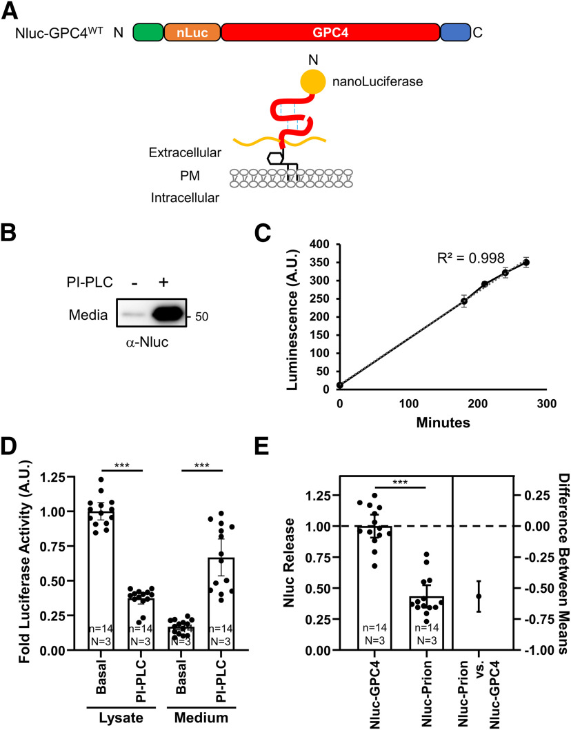
Luciferase assay for quantifying the release of GPC4 from astrocytes. A, Nluc is inserted at the N terminus, after the endogenous signal peptide, to preserve GPC4 trafficking. B, Primary astrocytes were nucleofected with Nluc-GPC4 and treated with and without PI-PLC. Western botting with α-Nluc antibody showed the expected 20 kDa size shift in the N-terminal fragment. PI-PLC treatment facilitates the release of Nluc-GPC4, confirming the GPI-anchorage of the construct. C, Representative trace of one experiment showing the linear kinetics of GPC4 release from astrocyte culture (R2 = 0.998). Error bars indicate the standard error of the mean. D, Astrocytes expressing Nluc-GPC4 were incubated in fresh media with and without PI-PLC for 3 h, and Nluc signal was measured in the cell lysate and media. Nluc signal was normalized to untreated lysate conditions for each biological replicate. PI-PLC treatment resulted in the decrease in the luciferase activity of the cell lysate and the corresponding increase in the activity in the media. These data show Nluc-GPC4 is quantitative in measuring released versus surface pools of GPC4. Error bars indicate 95% CI of the mean here and in following graphs. The requirement of the GPI-anchorage for PI-PLC-dependent release of GPC4 is shown in Extended Data Figure 2-1. E, The release rate (media over lysate activity) of Nluc-GPC4 and Nluc-Prion was normalized to Nluc-GPC4 release. Nluc-GPC4 is released ∼2-fold more than Nluc-Prion (t test p < 0.0001, Cohen’s d = 3.59); ***p < 0.001.

