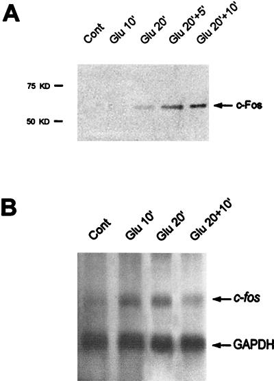
FIG. 1.
Glutamate-induced expression of c-Fos protein (A) and c-fos mRNA (B) in striatal slices. (A) Striatal slices were superfused with Krebs buffer alone (Cont) or in the presence of glutamate (100 μM) for 10 min (Glu 10′), 20 min (Glu 20′), or 20 min followed by 5 min (Glu 20′+5′) or 10 min (Glu 20′+10′) of Krebs buffer superfusion. At the end of the experiment, striatal slices were immediately lysed and processed for extraction of proteins. c-Fos protein expression was analyzed at the various time points by Western blotting with a specific anti c-Fos antibody. Fos protein is detectable at Glu20 and then increases at Glu20+5 and Glu20+10. (B) Total RNAs were extracted from the same striatal extracts (see Materials and Methods). c-fos and GAPDH mRNAs were detected on the same Northern blot. While GAPDH hybridization signals remain identical, c-fos mRNAs are induced at Glu10 and Glu20 and then their levels decrease slightly.

