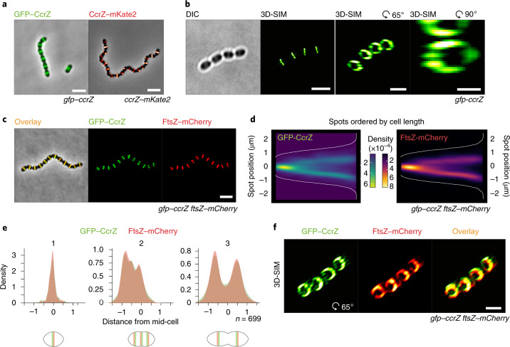
Fig. 2. CcrZ colocalizes with FtsZ at new division sites.
a, CcrZ localizes at mid-cell in live wild-type S. pneumoniae cells as observed by epifluorescence microscopy of GFP–CcrZ and CcrZ–mKate2. Scale bar, 3 µm. b, 3D-SIM of GFP–CcrZ and reconstructed volume projection (right) indicate that CcrZ forms a patchy ring at the mid-cell. Scale bar, 2 µm (left and middle right); 500 nm (right). c, GFP–CcrZ and FtsZ–mCherry colocalize in wild-type cells. Scale bar, 3 µm. d, Localization signal of GFP–CcrZ and FtsZ–mCherry in n = 699 cells of a double-labelled gfp–ccrZ ftsZ–mCherry strain, ordered by cell length and represented by a heatmap. e, GFP–CcrZ and FtsZ–mCherry colocalize during the entire cell cycle, as visualized when signal localization over cell length is grouped in three quantiles. f, 3D-SIM colocalization of GFP–CcrZ and FtsZ–mCherry shows a clear colocalizing ring with an identical patchy pattern. Note that for clarity, we did not correct for chromatic shift in the overlay. Scale bar, 2 µm.

