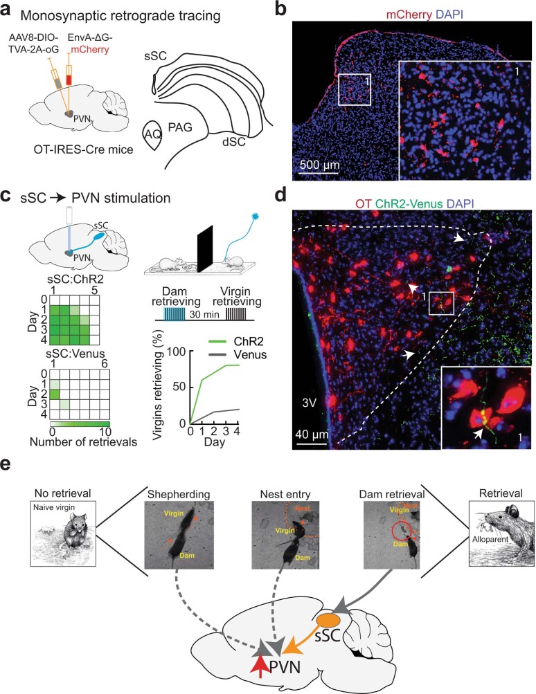
Extended Data Fig. 9. Superior colliculus input to PVN enhances acquisition of pup retrieval.
a, In Oxy-IRES-Cre virgins, we injected an AAV helper virus into PVN, followed two weeks later by injecting a rabies virus that carried mCherry fluorescent protein. Left, retrograde monosynaptic input tracing to OT-PVN neurons. Right, SC anatomy (AQ, aqueduct; PAG, periaqueductal gray; dSC, deep SC layers). b, We examined visual structures for mCherry expression (areas providing direct projections to OT-PVN cells). Immunohistochemistry showing mCherry+ cells (red) in the medial superficial layers of ipsilateral SC monosynaptically projecting to OT-PVN neurons. Blue, DAPI stain for cell number. Replicates from N=2 animals. c, Top, schematic of experimental design for optogenetic stimulation in PVN of fibres from ipsilateral sSC expressing ‘ChR2-Venus’ or ‘Venus’, during the ‘opaque barrier’ condition (‘Dam retrieving’ and ‘Virgin retrieving’, 10 pup retrieval trials per session; blue, optogenetic stimulation occurring during dam retrieval). Bottom left, individual performance on pup retrieval testing after each observation session. Bottom right, summary of behavioural data (Kaplan-Meier). d, Immunohistochemistry showing fibres from sSC (green, arrows) innervating PVN including near OT-PVN neurons (insert). Red, oxytocin neurons; green, Venus; blue, DAPI. 3V, third ventricle. Replicates from N=3 animals. e, Schematic of overall hypothesis connecting behavioural interactions between dam and virgin during co-housing to activation of PVN and OT-PVN neurons (red arrow).

