Fig. 2.
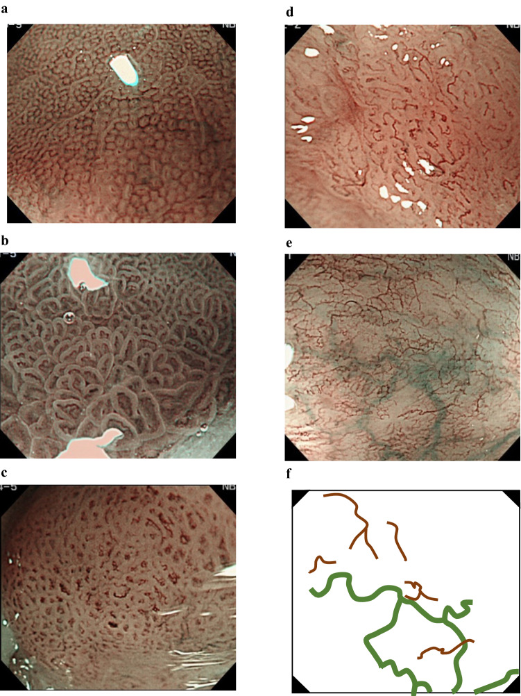
Representative endoscopic images using high-definition magnification NBI. a Regular mucosal pattern and regular vascular patterns. The mucosal pattern shows circular pits with similar sizes or forms arranged regularly. The vascular pattern demonstrates network-like vascular structure composed of spiral-like vessels located between pit-like mucosal patterns, and the vessel calibers change gradually. Histology from biopsy specimens showed fundic-type columnar epithelium including parietal cells and chief cells. b Regular mucosal pattern and regular vascular pattern. The mucosal pattern shows villous structures with density same as the surrounding area and clearly visible white zone with homogenous width. The vascular pattern located in the villous structure and the vessel calibers change gradually. Histology from biopsy showed cardiac-type columnar epithelium with specialized intestinal metaplasia and foveolar hyperplasia. c Irregular mucosal pattern and irregular vascular pattern. The mucosal pattern shows high-density villous patterns, and the vascular pattern demonstrates various forms with different calibers. Histology from endoscopic submucosal dissection showed well to moderately differentiated adenocarcinoma invading the lamina propria mucosae. d Invisible mucosal pattern and irregular vascular pattern. The vascular pattern shows irregularly bending and branching vessels. Calibers of the vessels change abruptly. Histology from endoscopic submucosal dissection showed well to moderately differentiated adenocarcinoma invading the muscularis mucosa. e Flat pattern with invisible mucosal pattern and regular vascular pattern. The flat pattern consists of a completely flat surface; normal-appearing, long branching vessels [brown lines in (f)]; and greenish thick vessels [bold green lines in (f)]. There is no demarcation line between completely flat area and the surrounding area. Histology of the biopsied tissue revealed tubular glands of specialized intestinal metaplasia that were covered by foveolar epithelium. f Schematic diagram of the endoscopic image shown in e

