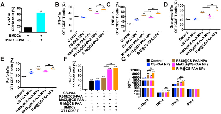
Figure 9.
R-M@CS-PAA NPs-treated DCs enhanced the cytotoxicity of OT-I CD8+ T cells to B16F10-OVA cells. (A) BMDCs were co-cultured with B16F10-OVA cells for 48 h, and the percentage of OVA+ BMDCs was detected by FCM. (B-F) In the presence of different NPs, BMDCs, OT-I CD8+ T cells, and B16F10-OVA cells were co-cultured for 48 h. The ratio of IFN-γ+ (B), TNF-α+ (C), granzyme B+ (D), and perforin+ (E) OT-I CD8+ T cells, and the proliferation of B16F10-OVA cells (F) were examined by FCM. (G) The expression of IL-12p70, TNF-α, IFN-β, and IFN-γ in the serum of mice treated with different NPs was detected by ELISA assay. *p < 0.05, **p < 0.01.

