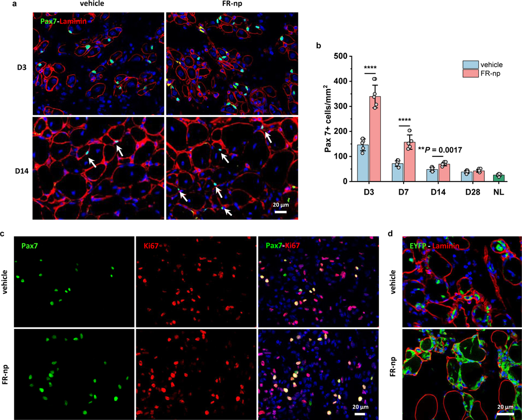
Fig. 8 |. Drug-loaded particles enhance muscle repair via promoting in situ satellite cell expansion.
a, Immunofluorescence analysis of Pax7+ cells in CTX-injured muscle treated with vehicle (np without drugs as a control, left panel) and FR-np (right panel) at day 3 (D3) and day 14 (D14) (n = 5 samples per group at each time point). b, Quantification of Pax7+ cells at different time points after treatment (n = 5 mice per group at each time point). NL represents the quantification of Pax7+ cells in normal adult TA muscle. c, Immunostaining of Pax7 (green) and Ki67 (red) in CTX-injured muscle treated with FR-np or vehicle at day 3 (n = 5 mice per group). d, Lineage-tracing of Pax7+ cells in CTX-injured muscle of Pax7-creER:Rosa26-EYFP mice. Image shows numerous EYFP+ cells around myofibers in FR-np-treated muscle at day 3 (n = 3 mice per group). Data are presented as mean ± SD. Two-tailed Student’s t-test. **P < 0.001, ****P < 0.0001.

