
FIGURE 3.
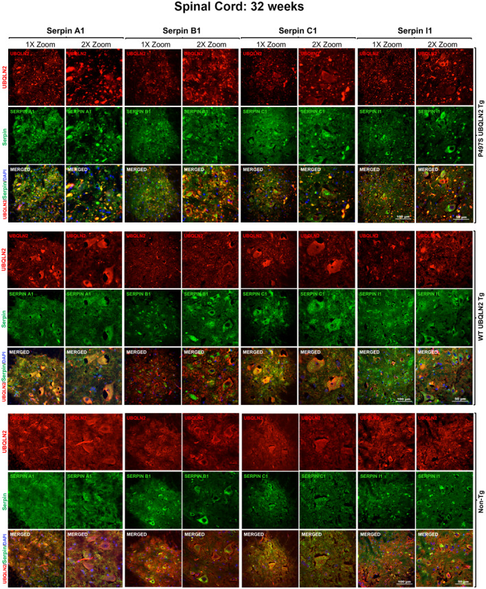
Colocalization of UBQLN2 and serpin proteins in puncta in the SC of P497S animals. Confocal microscopy images of the staining of UBQLN2 and Serpin A1, B1, C1, and I1 proteins and their corresponding merged image, including the DAPI panel, of the ventral horn region of the SC of 32‐week‐old P497S (top panel), WT356 (middle panel), and Non‐Tg (bottom panel) animals. Scale bars shown = 100 μm and 50 μm for 1× and 2× zoom images, respectively