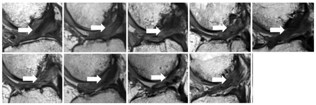
Figure 4:

Magnetic resonance imaging from 9 of the 10 patients in the scaffold enhanced repair group in the first-in-human study (sagittal view, 24 months after scaffold enhanced repair). All subjects had intact anterior cruciate ligament (ACL) fibers from the femoral to tibial attachment sites (arrows). The intact fibers have low signal intensity (black) reflecting highly organized tissue with little free water. (Used from Murray et al, OJSM, 201949).
