
Fig. 2. Intracerebral hemorrhage at 0.064T versus conventional imaging modalities (CT or 3T MRI).
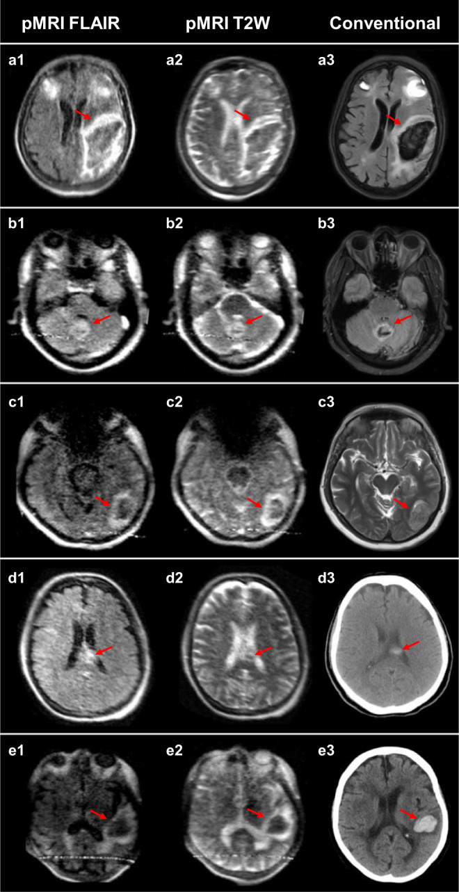
The first and second columns are low-field FLAIR and T2W images, respectively. The third column is a gold-standard clinical examination for comparison (3T MRI: a3, b3, c3, and e3; CT: d3). a Left isointense fronto-parietal intracerebral hemorrhage (ICH) with hyperintense rim and bilateral frontal hematomas. b Bilateral isointense cerebellar ICH with hyperintense rim. c Left hypointense occipital lobe ICH with hyperintense rim. d Left homogenous, hyperintense ICH in corpus collosum. e Left hypointense temporal ICH with hyperintense rim.