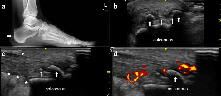
Fig. 6.
The Achilles tendon is among the most frequent sites of enthesopathic involvement in PsA. Forty-two-year-old male presenting with heel pain and difficulty walking. a Lateral X-Ray film shows a gross calcifications (thick arrow) at the distal Achilles tendon. These calficiations do not contact with the calcaneous bone and margins are ill-defined. On US examinations (b) these gross calcifications are identified as hyperechogenic surface with a posterior shadow (thick arrows) while other puntiforms hyperechogenic foci (thin arrow) do not have shadow and seems to be incipient calcification. c The Achilles tendon insertion is diffuse thickened (arrow head) and there is some fluid (*) at Kagger’s fat pad. d Color Doppler examination shows hypervascularity at the thickened Achilles tendon insertion at the calcaneous

