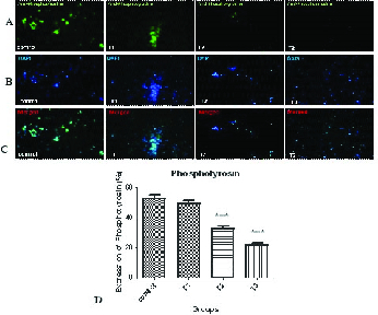
Figure 3.

Immunofluorescent staining of phosphotyrosine in the control and AA-treated groups (T1–T3). (a) Expression of phosphotyrosine antibody can be seen with a green color by the fluorescent microscope. (b) DAPI was added to examine the presence of sperm cells. The sperm nuclei were stained by blue color. (c) Combination of both protein expression and nucleus staining. (d) Comparison of the percentage of phosphotyrosine expression in different treatment groups.
