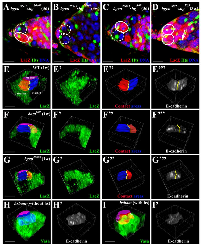
Figure 3.
The bgcn/bam-mediated GSC competition requires E-cadherin. In the germarial tips (A–D), the marked GSCs and the unmarked GSCs are highlighted by broken and solid circles, respectively. (A, B) Germarial tips harboring a three-day-old partial bgcn20915 shg10469 mutant GSC clone (A), and a three-week-old full mutant bgcn20915 shg10469 clone (B). (C, D) The germarial tips showing a three-day-old partial bgcn20093 shgR69 mutant GSC clone (C), and a recently lost three-week-old partial bgcn20093 shgR69 clone (arrow, D). (E–G) 3-D projections of germarial tips (pointing into the page) carrying control (E), bamΔ86 (F) and bgcn20093 (G) partial GSC clones, showing that the lacZ-negative (black) marked GSC (blue-contact area with cap cells colored purple) and the lacZ-positive (green) unmarked GSC (red-contact area) are separated by yellow lines. E′–G′ and E″–G″ show LacZ staining and the contact areas with cap cells for the marked GSC (red) and the unmarked GSC (blue), respectively. E‴–G‴ represent E-cadherin staining intensity in stem cell-niche junction. (H, I) The 3-D projections of a hs-bam germarial tip without (H) or with (I) heatshock treatments showing the stem cell-niche junction (H, blue; I, red) and E-cadherin accumulation in the junction (H′ and I′) between cap cells (purple) and GSCs (green, Vasa). The bars in A–D and in E–I represent 10μm and 5 μm, respectively.

