
FIGURE 3.
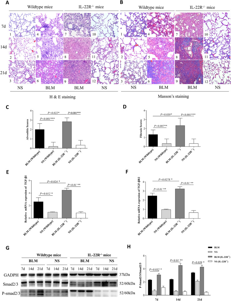
Elevated bleomycin (BLM)‐induced inflammation and fibrosis in interleukin (IL)‐22R−/− mice. Mice were intratracheally injected with 50 μl normal saline (NS) or BLM (5 mg/kg) to induce fibrosis at day 0. Each set of experiments was repeated three times. Six mice per group were used. (A1–6) The pathological changes of lung tissues on days 7, 14, and 21 in NS and BLM treated wild‐type (WT) mice by HE staining (×200). (A7–12) The pathological changes of lung tissues on days 7, 14, and 21 in BLM and NS treated IL‐22R−/ − mice by HE staining (×200). (B1–6) The pathological changes of lung tissues at day 7, 14, and 21 days in NS and BLM treated WT mice by Masson's trichrome (MS) staining (×200). (B7–12) The pathological changes of lung tissues on days 7, 14, and 21 in BLM and NS treated IL‐22R−/‐ mice by MS staining (×200). (C) Comparison of the pathological scores of alveolitis at 7 days after treatment with BLM in WT mice and IL22R−/ − mice. (D) Comparison of the pathological scores of fibrosis at 21 days after treatment with BLM in WT (n = 6) and IL22R−/ − mice (n = 6). (E–F) The mRNA expressions of transforming growth factor (TGF)‐β1 and TGF‐βR2 in the lungs of BLM, NS treated WT and IL22R−/‐ mice at 21 days. (G–H) The protein expressions of P‐smad2/3 in the lung tissues of BLM, NS treated WT, and IL22R−/‐ mice by western blot analysis