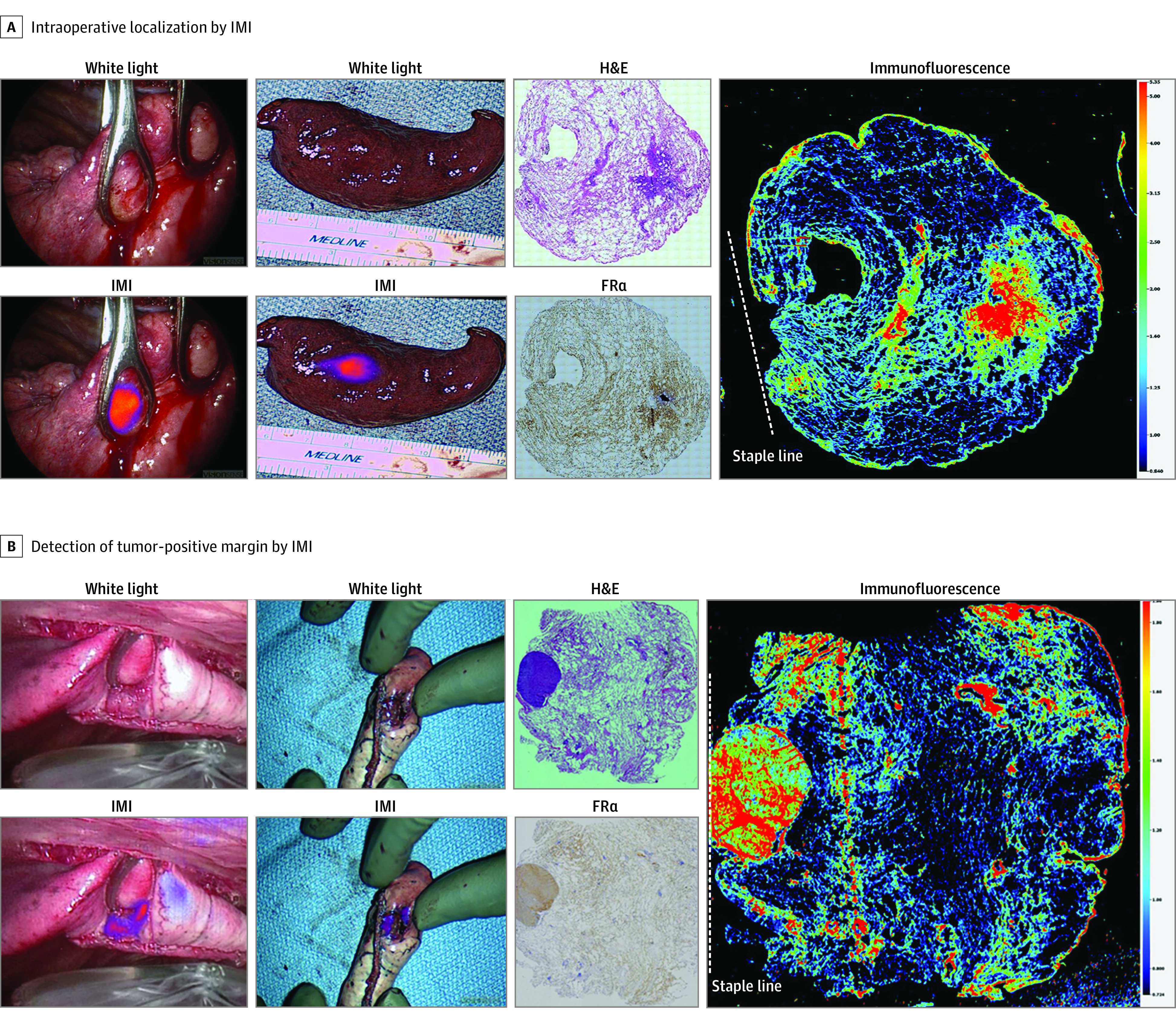
Figure 4. Intraoperative Molecular Imaging (IMI) Alters Surgical Management of Nonpalpable Lesions.

A, Representative images from a tumor that was not able to be detected by traditional surgical methods of visual inspection and palpation. IMI clearly identifies the lesion of interest both in situ and ex vivo. Histologic evaluation using hematoxylin-eosin (H&E) staining, folate receptor alpha (FRα) staining, and immunofluorescence confirm that fluorescence is concentrated in the area of tumor, which overexpressed FRα and was far from the staple line (dashed line). B, Identical images of a nonpalpable tumor with a tumor-positive margin as indicated by fluorescence on the staple line on ex vivo imaging and confirmed by immunofluorescence microscopy.
