Figure 6.

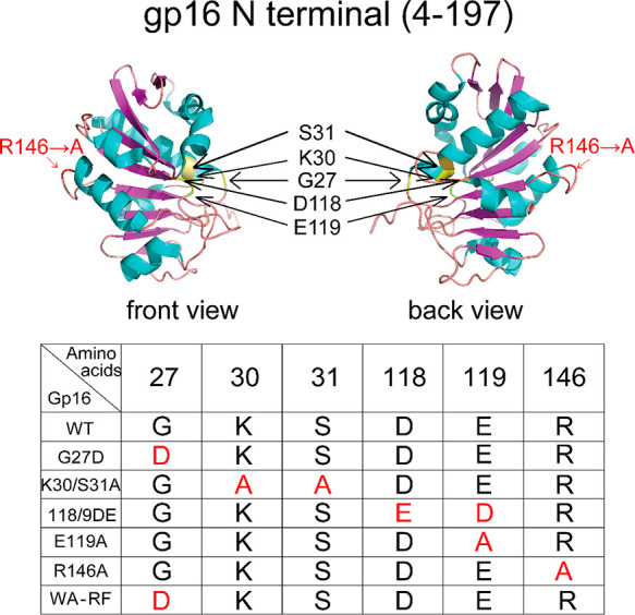
Crystal structure of the N terminus of gp16 (top figure, PDB file: 5HD9) and the location of the mutations (bottom table). The red amino acids in the table indicate the mutation sites.

Crystal structure of the N terminus of gp16 (top figure, PDB file: 5HD9) and the location of the mutations (bottom table). The red amino acids in the table indicate the mutation sites.