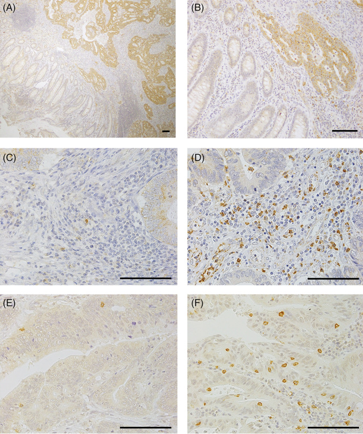
FIGURE 1.

Immunohistochemistry for LOX‐1 and CD8 in CRC tissues. A and B, LOX‐1+ cells were localized in the tumor and stroma (A, magnification ×40; B, ×200). C, Low expression of LOX‐1. D, High expression of LOX‐1. E, Low density of CD8+ CTL. F, High density of CD8+ CTL. Scale bar = 100 μm. CRC, clinical colorectal cancer; LOX‐1, lectin‐like oxidized low‐density lipoprotein receptor‐1; CD8+ CTL, CD8+ cytotoxic T‐lymphocytes
