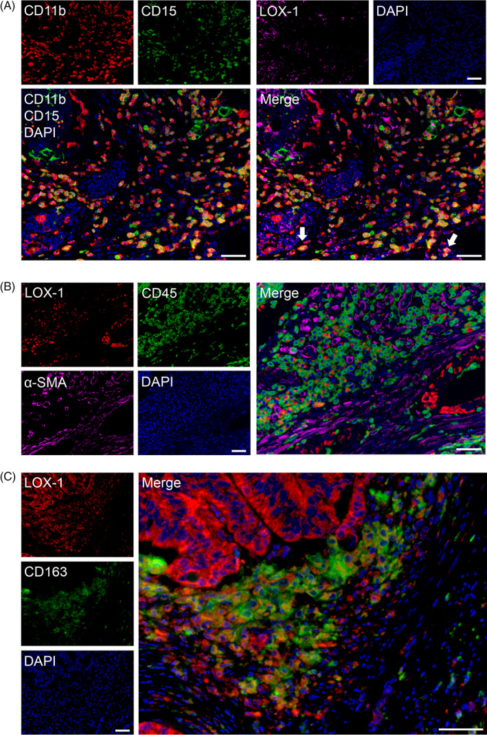
FIGURE 4.

Evaluation of LOX‐1+ cells using multiplex immunofluorescence in CRC tissues. A, MDSC markers, CD11b (red), CD15 (green), and LOX‐1 (magenta). Cells co‐expressing CD11b+ CD15+ are in yellow. Some LOX‐1+ stromal cells partially express CD11b+ and CD15+ cells (white arrows). B, LOX‐1+ stromal cells expressed CD45 (green) but did not express α‐SMA (magenta). C, Almost LOX‐1+ cells expressed CD163 (green). Nuclei were stained with DAPI (blue). Scale bar = 50 μm. CRC, clinical colorectal cancer; LOX‐1, lectin‐like oxidized low‐density lipoprotein receptor‐1; MDSCs, polymorphonuclear MDSCs
