Figure 5.

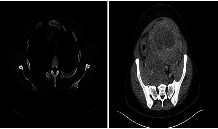
A staging CT chest, abdomen and pelvis showed a large mediastinal lymph node in the anterior mediastinum (8 mm) and a right pelvic nodal mass lesion (120×65 mm). A mixed solid cystic mass lesion within the uterus was observed.

A staging CT chest, abdomen and pelvis showed a large mediastinal lymph node in the anterior mediastinum (8 mm) and a right pelvic nodal mass lesion (120×65 mm). A mixed solid cystic mass lesion within the uterus was observed.