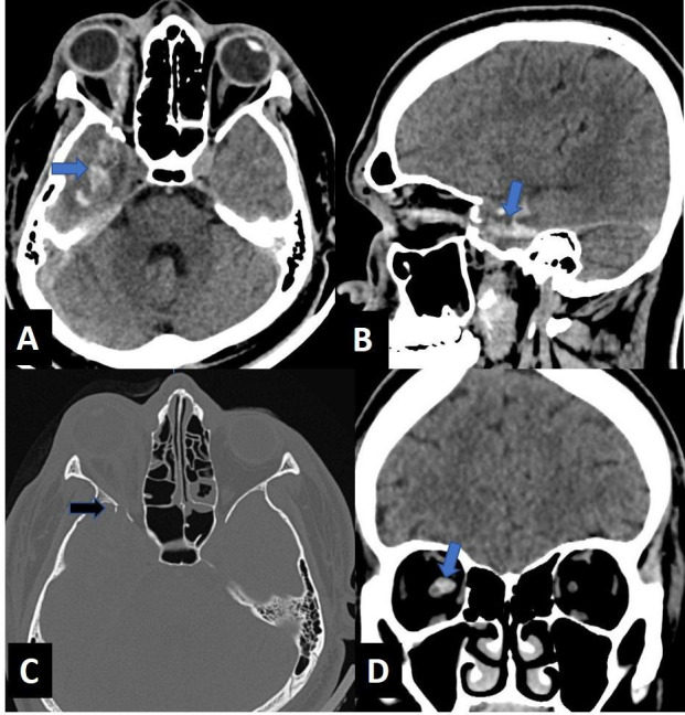
Figure 1.

Axial (A) and sagittal (B) images of the head reveal a linear hyperdense track (curved arrow), which is passing medial to lateral, entering the right orbit medial to the globe along the medial canthus, involving the intraconal compartment with intracranial extension causing temporal lobe laceration (blue arrow). Axial (C) bone window image of the orbit reveals displaced fracture of the right greater wing of sphenoid with few tiny fracture fragments seen protruding into the intracranial extra-axial region (black arrow). Coronal (D) image of the orbit reveals the hyperdense tract closely abutting the right optic nerve (blue arrow).
