
Figure 2.
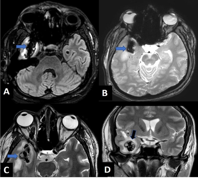
Axial FLAIR (A), gradient recalled echo (B), T2WI (C) and coronal T2WI (D) images revealing linear hypointense tract (blue arrow) showing blooming on gradient recalled echo images and laceration involving the right temporal region (black arrow). FLAIR, Fkuid-attenuated inversion recovery; T2WI, T2 weighted image.