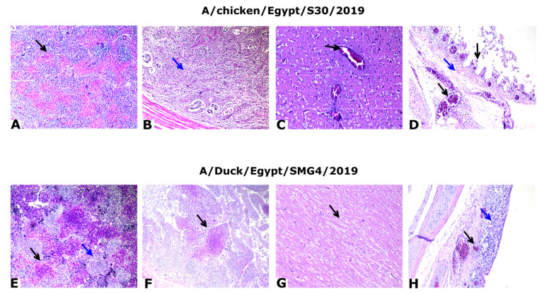
Figure 5.
Histopathology slices from the tissue of infected chickens. (A) Spleen of A/chicken/Egypt/S30/2019(H5N8) at 2 dpi showing severe multifocal necrotic tissue (black arrow); H&E, ×100. (B) Cecal tonsils at 2 dpi showing mild depletion of lymphocytes (star); H&E, ×100. (C) Cerebrum of control positive at 3 dpi showing congested blood vessels (black arrow); H&E, ×200. (D) Trachea of control positive at 2 dpi showing sloughed epithelium, edema (blue arrow), and congested blood vessels in lamina propria (black arrow); H&E, ×200. (E) Spleen of A/Duck/Egypt/SMG4/2019(H5N8) at 2 dpi showing severe depletion of lymphocytes (blue arrow) with multifocal necrotic areas (black arrow); H&E, ×100. (F) Cecal tonsils at 2 dpi showing multifocal necrotic patches (black arrow); H&E, ×100. (G) Cerebrum at 2 dpi showing demyelination (black arrow); ×200. (H) Trachea at 2 dpi showing thickening of the wall of the mucosal layer due to congested blood vessels (black arrow), edema, and mononuclear cell infiltration (blue arrow); H&E, ×200.

