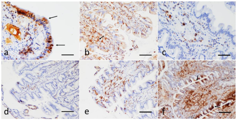
Figure 2.
Sections of the duodenum from dogs with IRE. Immunohistochemical evaluation of the epithelial and the lamina propria infiltrating lymphocytes; P-gp antibody immunohistochemistry, Streptavidin-biotin immunoperoxidase technique (brown stain). (a) Focal, strong cytoplasmic expression of P-gp in enterocytes, reinforced on the apical luminal portion (arrows), scale bar: 100 µm. (b) Diffuse, mild, with focal expression at the tips of the villi. Note the strong positivity also in numerous submucosal lymphocytes (arrow), scale bar: 300 µm. (c,d) Infiltration of the duodenal lamina propria with a scarce number of P-gp positive lymphocytes (score 2). There is no expression of P-gp at the enterocytes; scale Bar: 100 µm. (e) Strong P-gp positivity in a moderate number of laminae propria lymphocytes (score 3); scale bar: 300 µm. (f) Strong and diffuse P-gp expression in the lymphocyte submucosal infiltrate (score 4). Note the marked focal positivity also in the apical portion of the duodenal epithelial cells (arrows); scale bar: 300 µm.

