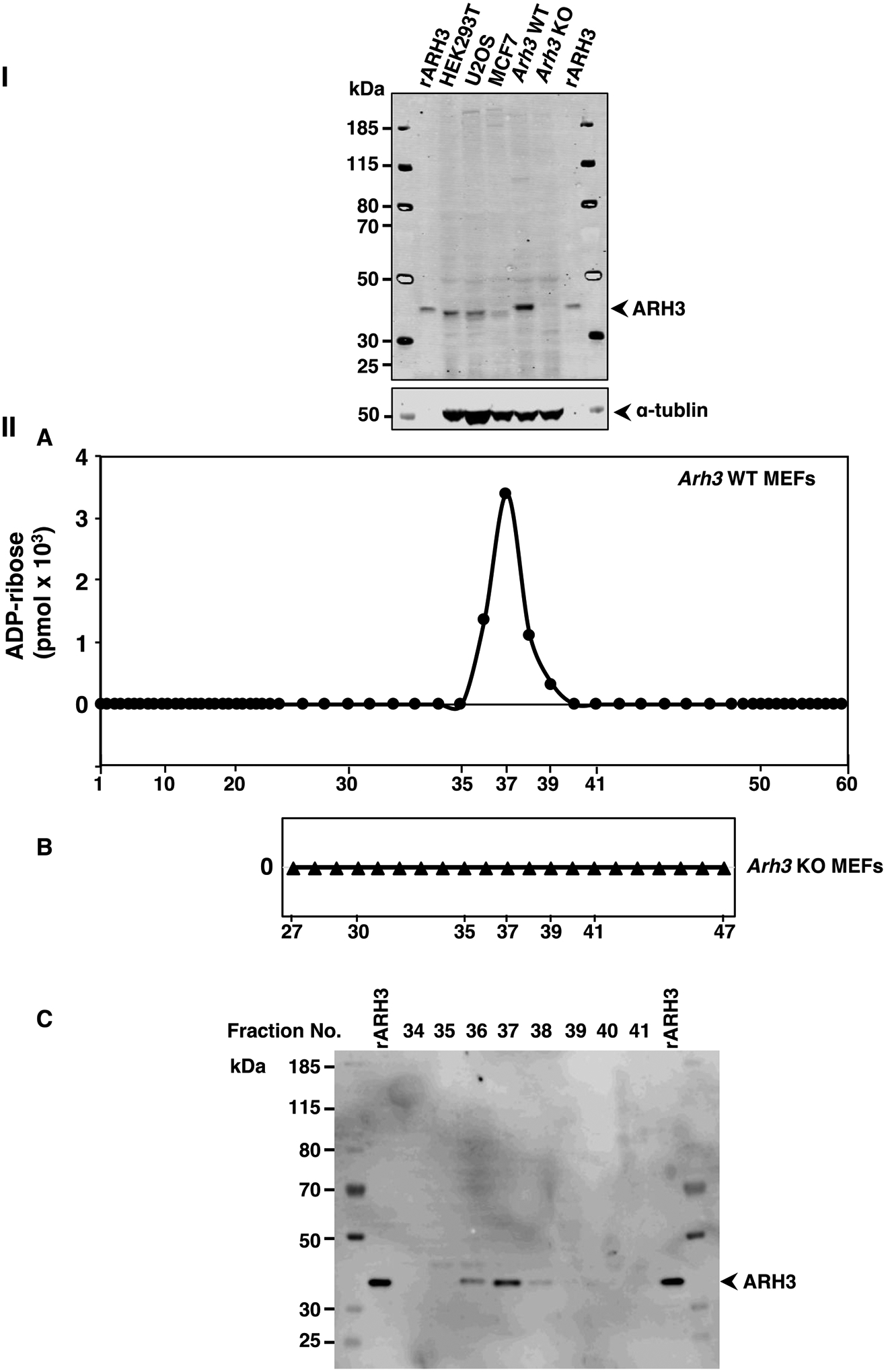
Figure 2.

ARH3 expression and α-NADase activity in lysates from wild-type (WT) and Arh3−/− MEFS.
Western blot analysis of ARH3 expressed in whole cell lysates (80μg) from HEK293T, U2OS, MCF7, WT MEFs and Arh3−/− MEFs compared to recombinant ARH3 (see Methods). The blot was incubated with fluorescence-labeled secondary antibody to quantify the bands, normalized to tubulin for detection using the Odyssey Infrared Imaging System and repeated twice. Immunoreactivity of WT MEFS>>U2OS ≅ HEK293T>rARH3>MCF7>>> Arh3−/− MEFs (I). ADP-ribose (pmols) detected in fractions (180μl) of WT MEF lysate (612μg) or Arh3−/− MEF lysate (666μg) separated by weak anion-exchange HPLC (see Methods) with fractions that were incubated with α-NAD+ (50μM), MgCl2 (10mM) in Tris-HCl pH 7.5 (50mM) overnight at 30 °C (IIA,B). Data represent weak anion-exchange separations and α-NADase analysis repeated twice. Western blot analysis of ARH3 expression in fractions (20μl) separated by weak anion-exchange HPLC (IIA) detected by the Odyssey Infrared Imaging System and repeated twice (IIC).
