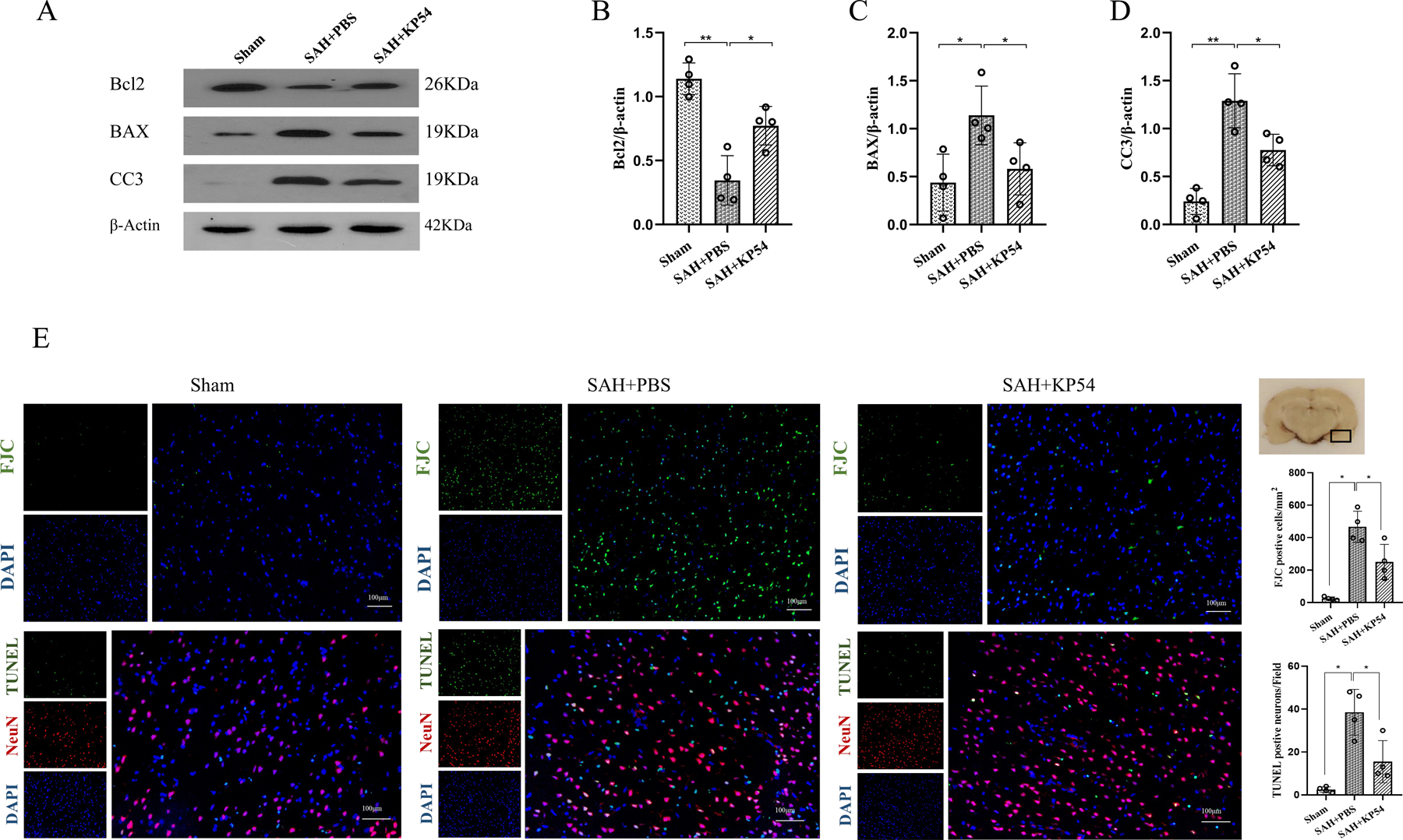
Fig. 5. KP54 treatment attenuated the neuronal damage at 24 h after SAH.

(A) Representative western blot bands of the brain apoptosis markers at 24 h after SAH; (B-D) Densitometric quantification for the apoptosis markers in brain at 24 h after SAH; (E) Representative microphotographs and quantitative analysis for FJC and TUNEL staining in the ipsilateral cortex of rat brain. The small black rectangular box in the coronal brain slice (the right top panel) indicates the area for staining quantification. Scale bar = 100 μm. *P < 0.05, **P < 0.01. Data were represented as mean ± SD, n = 4 per group, one-way ANOVA Tukey test.
