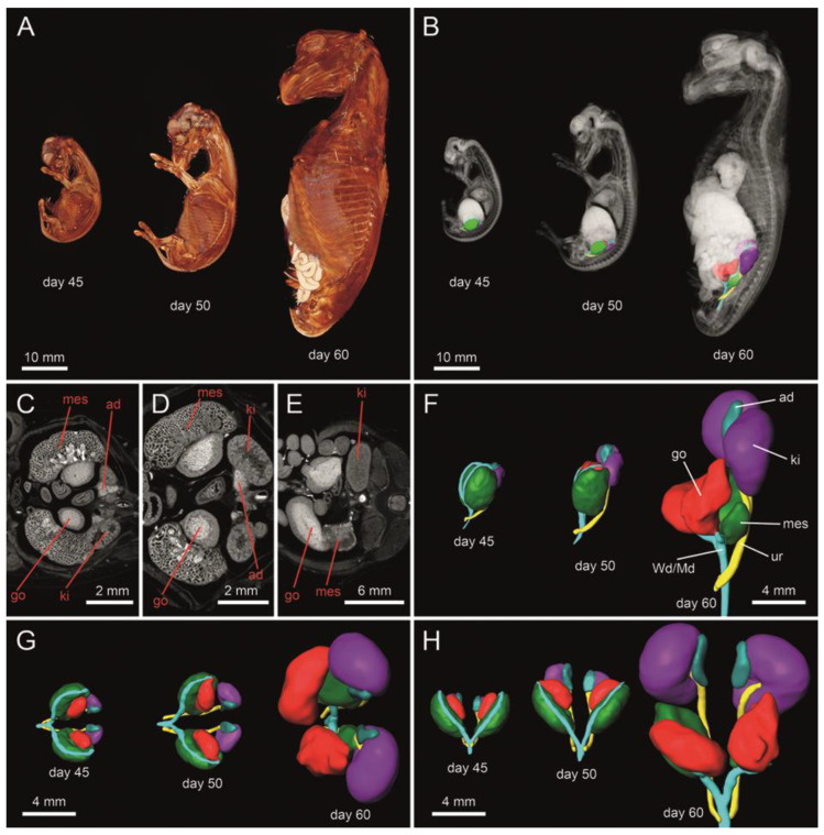
Figure 1.
The 3D reconstruction of female equine fetuses at 45, 50 and 60 days of gestation (A). Structural representation of the urogenital tract in transverse section (C–E). 3D architecture of the urogenital tract (B,F–H) in situ (B), as well as from lateral (F), cranial (G) and ventral view (H). A well identifiable delimitation between the cortical and the medullar region can be observed in the developing gonads (C–E). Color-coded abbreviations: go, gonad (red); Wd/Md, Wolffian (mesonephric) duct/Müllerian (paramesonephric) duct (turquois green); ad, adrenal gland (blue); ki, kidney/metanephros (violet); mes, mesonephros (green); ur, ureter (yellow).

