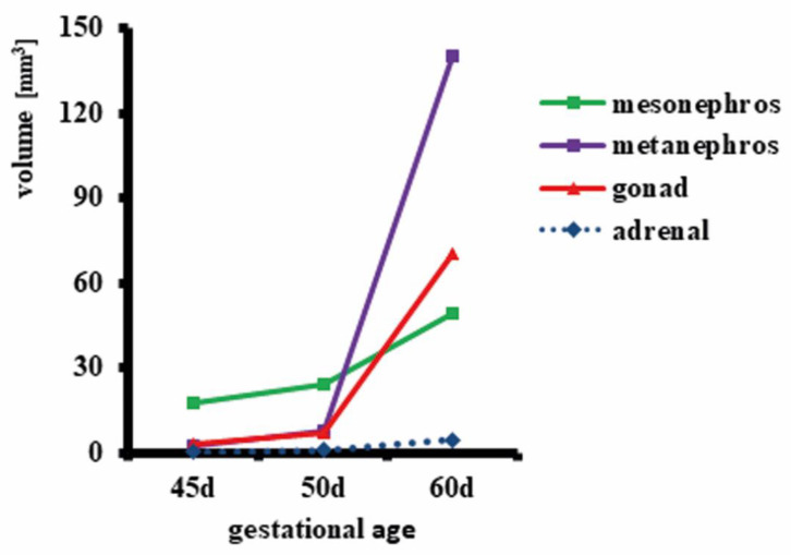
Figure 2.
Size (volume) of mesonephros, metanephros, gonads and adrenal glands in female horse fetuses at 45, 50 and 60 days of gestation. After microCT scanning of one female fetus from each stage, image volumes were imported into Amira 6.4 and the urogenital region (mesonephros, metanephros, gonads, and adrenal glands) was divided using manual digital segmentation tools. Surface models were created from segmentation masks and volumes calculated by the software.

