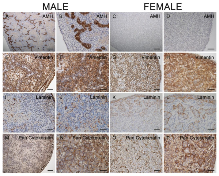
Figure 4.
Immunostaining for AMH (A–D), vimentin (E–H), pan-cytokeratin (I–L) and laminin (M–P) in male (A,B,E,F,I, J,M,N) and female (C,D,G,H,K,L,O,P) gonads from 60-day-old horse fetuses. AMH was only expressed in the male fetal testes (A,B). Vimentin was expressed in interstitial cells of gonads from both sexes (E–H). The surface epithelium of fetal male and female gonads, as well as the cord-like structures in females, stained positive for cytokeratins (I–L). A basal lamina-like structure surrounded the developing seminiferous tubules in males and the cord-like structures in females (M–P). (B,D,F,H,J,L,N,P) represent higher magnifications of their left images. Scale bars for all pictures: 50 µm.

