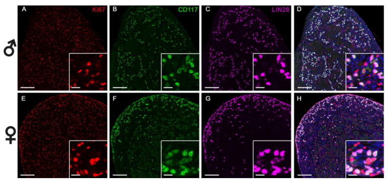
Figure 7.
Triple immunofluorescence staining of fetal male (A–D) and female (E–H) horse gonads at 60 days of gestation. Proliferation marker Ki67 (red) is present throughout the gonad in both sexes and has a different localization than LIN28 and CD117. A distinct staining pattern depending on sex is observed for CD117 (green) and LIN28 (pink): in male gonads, CD117 and LIN28 are scattered throughout the gonad, while in females they are mainly localized at the periphery of the gonad. Scale bar: 100 µm for large pictures, 25 µm for the inserts. Blue staining denotes cell nuclei.

