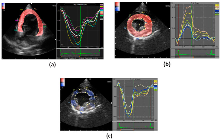
Figure 3.
An example of strain analysis of the LV in a healthy dog; (a) longitudinal strain (apical 4-chamber view); (b) radial and (c) circumferential strains (parasternal short axis view). In longitudinal and circumferential strains, the deformation is illustrated by a negative curve during systole, whereas in radial strains it is illustrated by a positive curve.

