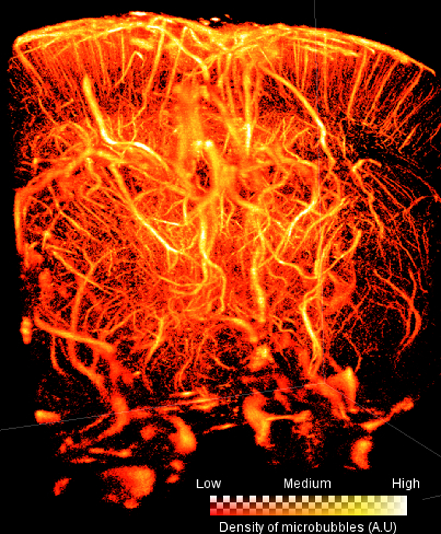
Figure 10:

Volumetric Ultrasound Localization Microscopy implemented on an anesthetised rat brain using a 2D matrix array with center frequency 9MHz (Heiles et al. 2018). Voxel size is 10 μm, region of imaging is 13.28mm deep, 10.5mm wide, and 11.4mm in length. The “glow” colormap was used to encode microbubble density. The brighter the color of the voxel, the more microbubbles have passed through that voxel.
