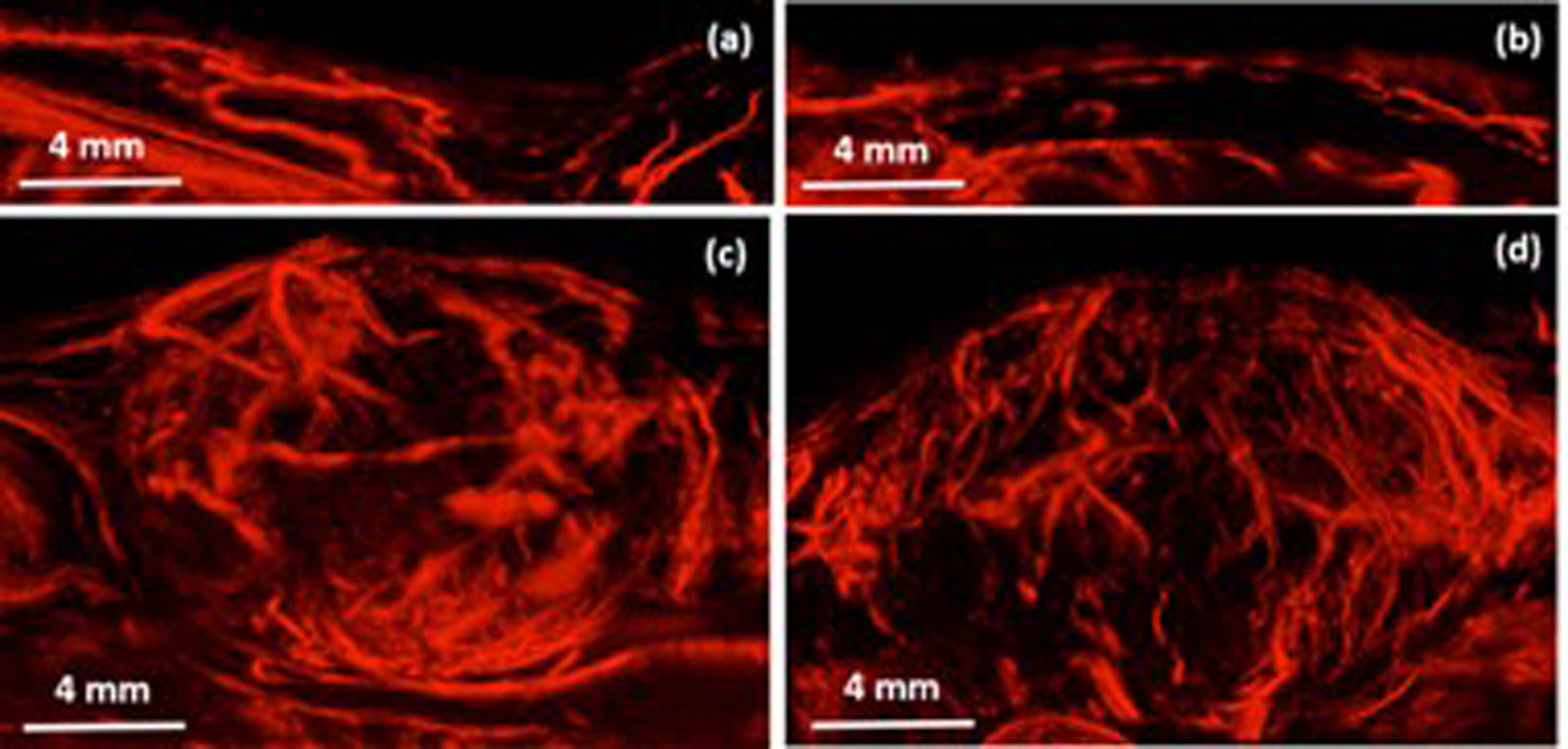
Figure 5.

Maximum intensity projections of 3D super-resolution imaging of healthy microvasculature (a-b) and tumor associated microvasculature (c-d). Smallest vessels resolved were approximately 25 microns in diameter, approximately 6x improved from achievable with acoustic angiography at a similar depth. Reproduce with permission from: F. Lin, S. E. Shelton, D. Espindola, J. D. Rojas, G. Pinton, and P. A. Dayton, “3-D Ultrasound Localization Microscopy for Identifying Microvascular Morphology Features of Tumor Angiogenesis at a Resolution Beyond the Diffraction Limit of Conventional Ultrasound,” Theranostics, vol. 7, no. 1, pp. 196-204, 2017.
