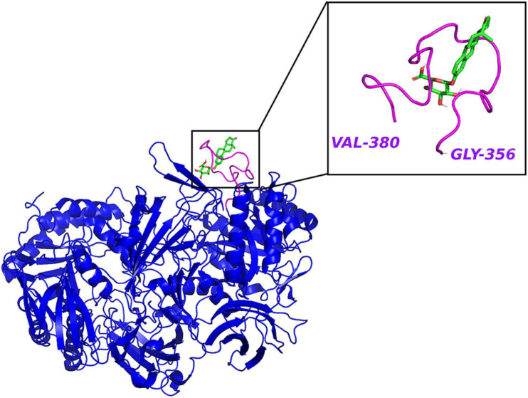
FIGURE 3.
The crystal structure of E. coli β-glucuronidase (PDB ID: 3LPF) with estrone-3-glucuronide (PubChem CID 115255) docked in AutoDock Vina (Trott and Olson, 2010) with affinity –7.2 kcal/mol. Hydrogen atoms were added to the enzyme structure. The default docking protocol was applied and the possible poses were saved. The view of the docking results and analysis of their surface with graphical representations were done using PyMOL 2.4 (Schrodinger, 2015).

