Figure 6.
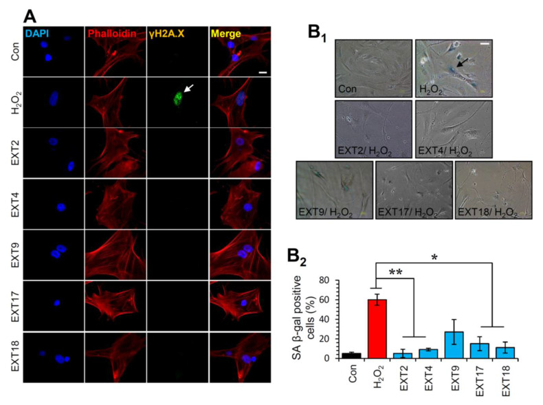
Protection of cells against oxidative stress-mediated premature senescence by the extracts. (A) Immunofluorescence images following detection of the phosphorylation form of H2A.X (γH2A.X) on Ser139 in BJ cells incubated with the indicated extracts for 24 h; cells nuclei were counterstained with DAPI and Phalloidin. (B1) Representative light field images following SA β-gal staining of control BJ cells or cells treated (three exposures of 48 h each) with 300 μM H2O2 in the presence or absence of the extracts. (B2) Relative (%) number of SA β-gal positive cells (mean of 10 optical fields) following treatment of cells exactly as in (B1). Bars ± SD. * p < 0.05; ** p < 0.01. Bars, in (A) 10 μM and in (B1) 100 μM.

