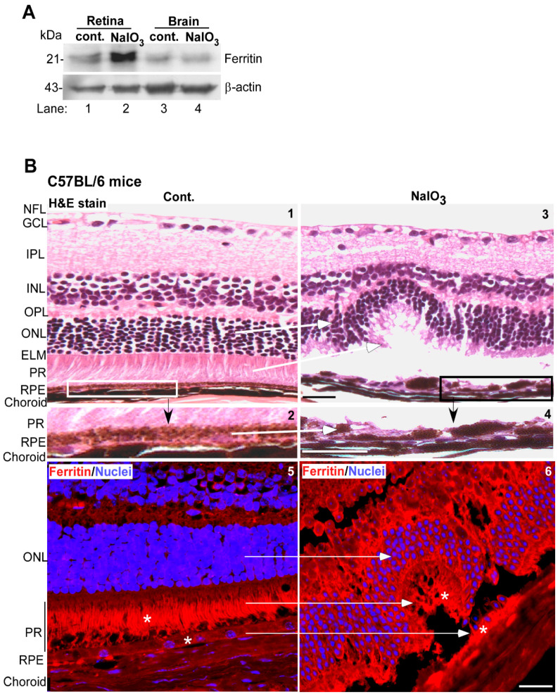
Figure 5.
NaIO3 upregulates ferritin and disrupts retinal cell layers in mouse models. (A) Probing of retinal and brain lysates from NaIO3 and control mice for ferritin showed significantly higher levels in retinal samples (lane 2 vs. 1) relative to controls. Brain samples from the same mice showed a similar level of ferritin (lane 4 vs. 3). (Tissue from 5 mice was pooled for this experiment). The membranes were re-probed for β-actin as a loading control. Full blots and their details are provided in Supplementary Figure S1. (B) Hematoxylin and Eosin (H&E) staining of mouse retina showed well-defined layers in control mice (panel 1). The boxed area is enlarged in panel 2. Mice administered NaIO3 showed disruption of the RPE and photoreceptor cell layers (panel 3). Scale bar: 25 µm. The boxed area is enlarged in panel 4. Scale bar: 25 µm. Immunoreaction for ferritin showed increased expression in the RPE and photoreceptor cell layers of NaIO3-treated mice relative to controls (panel 6 vs. 5, marked by *). Scale bar: 25 µm. A serial section treated with rabbit IgG and Alexa Fluor 546-conjugated secondary antibody (red) showed no reaction (Supplementary Figure S3). RPE: retinal pigment epithelium; PR: photoreceptors; ELM: external limiting membrane; ONL: outer nuclear layer; OPL: outer plexiform layer, INL: inner nuclear layer, IPL: inner plexiform layer, GCL: ganglion cell layer; NFL: nerve fiber layer.

