Figure 6.
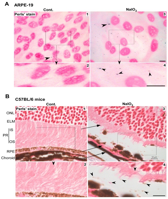
Iron-loaded vesicles are released in the extracellular milieu by NaIO3 treatment. (A) Staining with Perls’ reagent showed several blue dots intracellularly and extracellularly in NaIO3-treated ARPE-19 cells (panel 3, arrowhead). Scale bar: 10 µm. Boxed area is enlarged in panel 4. Scale bar: 10 µm. A rare blue dot was also noted in control cells (panel 1, arrowhead). Boxed area is enlarged in panel 2. Scale bar: 10 µm. (B) Perls’ reaction of retinal sections from NaIO3-treated mice showed a positive reaction in the space between RPE and PR cells (panel 3, arrowhead), better visualized in the enlarged boxed area (panel 4, arrowheads). Scale bar for panel 3: 10 µm. Scale bar for panel 4: 10 µm. RPE: retinal pigment epithelium; PR: photoreceptors; OS: outer segment of photoreceptors; IS: inner segment of photoreceptors; ELM: external limiting membrane; ONL: outer nuclear layer.

