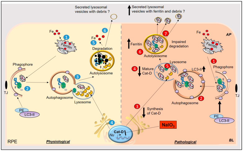
Figure 7.
Graphical representation of NaIO3-induced model of AMD in-vitro and in-vivo. Physiological: (1) Iron-loaded ferritin is transported to the phagophore by NCOA4, (2) for degradation with the rest of the cell debris. Levels of LC3II increase with the maturation of phagophore to autophagosome. (3) Autophagosome fuses with lysosomes, (4) where lysosomal enzymes, including cat-D, degrade ferritin and cellular debris. (5) Iron from ferritin is released in the cytosol to be utilized for metabolic purposes. (6) Other debris undergoes autolysis or is secreted via an unconventional lysosomal secretory pathway to the extracellular milieu. Pathological: (1) In NaIO3-induced AMD, iron-loaded ferritin is delivered to the phagophore, (2) which matures into autophagosomes. Levels of LC3II are increased because of accumulation of debris in autophagosomes. (3 and 4) Synthesis and maturation of cat-D are impaired by NaIO3, (5) resulting in the accumulation of iron-loaded ferritin in lysosomes and (6) autolysosomes. (7) Iron-loaded ferritin is secreted in lysosomal vesicles in the extracellular milieu, which is probably internalized by the adjacent photoreceptors, resulting in iron-mediated oxidative stress.

