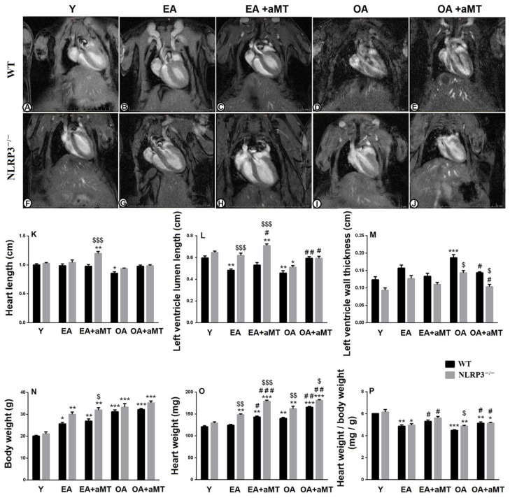
Figure 1.
Impact of NLRP3 deficiency and melatonin therapy on cardiac magnetic resonance imaging and anthropometric parameters during aging. (A–E) Magnetic resonance imaging of the heart of young (Y), early-aged (EA), early-aged with melatonin (EA + aMT), old-aged (OA), and old-aged with melatonin (OA + aMT) WT mice. (F–J) Magnetic resonance imaging of the heart of Y, EA, EA + aMT, OA, and OA + aMT NLRP3−/− mice. (K) Analysis of the heart length (cm). (L) Analysis of the luminal length of the left ventricle (cm). (M) Analysis of the thickness of the left ventricular wall (cm). (N) Analysis of the body weight (g). (O) Analysis of the heart weight (mg). (P) Analysis of the ratio of the heart weight to the body weight (mg/g). * p < 0.05, ** p < 0.01 and *** p < 0.001 vs. Y; # p < 0.05, ## p < 0.01 and ### p < 0.001 vs. aged group without melatonin; $ p < 0.05, $$ p < 0.01 and $$$ p < 0.001 vs. WT mice.

