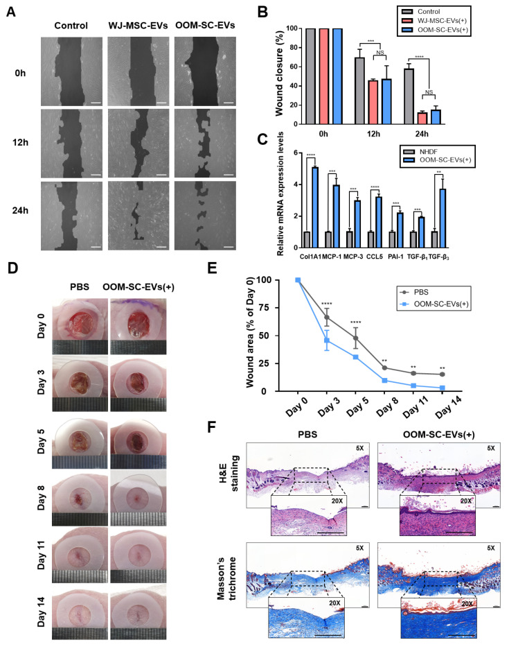
Figure 6.
In vitro and in vivo wound healing capacities and the anti-wrinkling activity of the OOM-SC-EVs (A) Effect of OOM-SC-EVs in the closure of in vitro scratches of fully confluent NHDFs tablet 10. μg/mL mitomycin C for 2 h and then scratched with a 200-μL tip. Subsequently, cells were exposed to 1.5 × 10⁹ particles of OOM-SC-EVs or WJ-MSC-EVs in a time-dependent manner (0, 12, and 24 h). Scale bar = 200 μm. (B) Graphic diagram representing the in vitro scratch assay in Figure 6A, and the wound closure was evaluated using TScratch software. Data are presented as mean ± SD. Statistical significance was determined using RMANOVA with post-hoc analysis: *** p < 0.001, and **** p < 0.0001, NS; not significant. (C) RT-PCR results showing changes in the expression levels of anti-wrinkle-associated genes. OOM-SC-EV treatment markedly increased the expression levels of collagen synthesis-related genes, namely ColA1, MCP-1, MCP-3, CCL-5, PAI-1, TGF-β1, and TGF-β3, which are related to high collagen synthesis and the treatment of wrinkles. Data are presented as mean ± SD. Statistical significance was determined using Two-tailed t test: ** p < 0.01 *** p < 0.001, and **** p < 0.0001. (D) In vivo wound healing assay. In this model, two full-thickness skin wounds, on the back of each mouse, were excised via a sterile biopsy punch, followed by the subcutaneous injection of 1.5 × 10⁹ particles/mL OOM-SC-EVs diluted in 30 μL PBS. Wound closure was monitored every two days until day 14. (E) Graphical data depicting the closure of the wound area as calculated relatively to the original wound area on day 0 after inducing the injury (n = 5) and in a time-dependent manner. Data are presented as mean ± SD. Statistical significance was determined using RMANOVA with post-hoc analysis: ** p < 0.01, and **** p < 0.0001. (F) Hematoxylin and eosin and Masson’s trichome staining of the mice skin after sacrifice (on day 14). Two weeks after injection of OOM-SC-EVs into the experimental wound, the skin tissues were fixed with 4% PFA and then dehydrated using various concentrations of alcohol, followed by paraffin embedding. For the evaluation of regeneration and re-epithelization of the wound after OOM-SC-EV application, the sections were stained with hematoxylin and eosin. Further, Masson’s trichrome staining was performed to estimate collagen synthesis rate. Scale bar = 200 μm.

