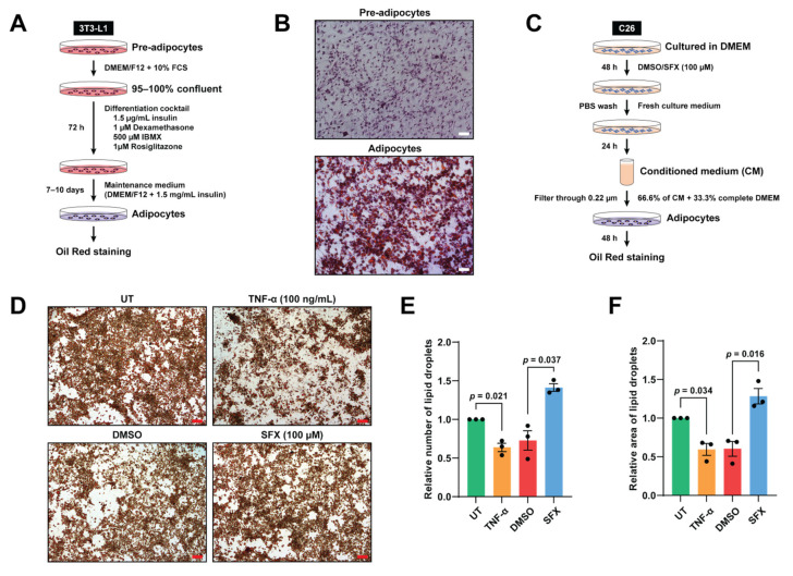
Figure 3.
SFX reduces the loss of lipid droplets induced by C26 conditioned medium (CM) in vitro. (A) Schematic representation of the differentiation of pre-adipocytes to adipocytes using differentiation and maintenance medium. (B) Cells that were fully differentiated into adipocytes were stained with Oil Red O. First column: undifferentiated pre-adipocytes. Second column: differentiated adipocytes showing lipid droplets under a phase contrast microscope (purple staining represents hematoxylin). Magnification of 10×, scale bar: 200 µm. (C) Schematic representation of the cell culture, drug treatment and collection of CM. C26 cancer cells were cultured in DMEM medium with SFX (100 µM) or vehicle alone for 48 h. Following PBS wash, C26 cancer cells were incubated in DMEM complete medium for 24 h to produce CM. (D) Oil Red O lipid staining under a phase contrast microscope upon incubation with DMEM complete medium (untreated), 100 ng/mL TNF-α (positive control), C26 SFX, or vehicle alone treated C26 CM treatment for 48 h. Magnification of 10×, scale bar: 200 µm. (E,F) Dot plots representing quantification of the relative total number and area of lipid droplets, respectively, using ImageJ. All data are representing mean ± SEM. Statistical significance was examined by paired two-tailed Student’s t-test, n = 3 biologically independent experiments.

