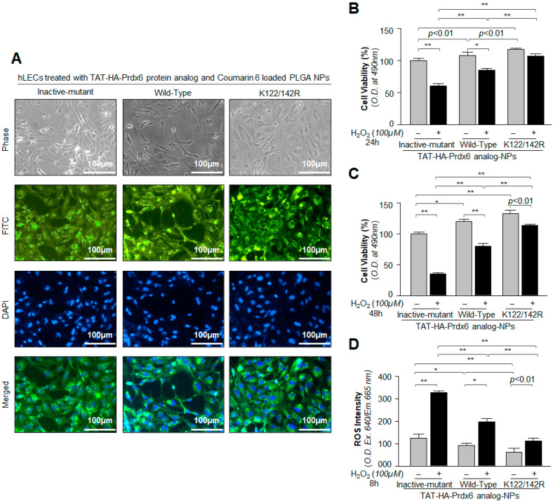
Figure 4.
Analysis of Prdx6 analog-loaded PLGA-NPs cellular internalization and evaluation of cytoprotective potential against H2O2-induced oxidative cell death. (A) A representative of photomicrographs displaying the internalization of Prdx6 analog-loaded PLGA-NPs (with Coumarin-6, a fluorescence marker) in hLECs as indicated. (B–D) hLECs treated with Sumoylation-deficient protein TAT-HA-Prdx6K122/142R-NPs engendered higher resistance against oxidative stress than TAT-HA-Prdx6WT-NPs. hLECs were incubated with TAT-HA-Prdx6WT and its mutant proteins loaded NPs for 6h as indicated; then, the cells were tryspinized and harvested on coverslips to take photomicrographs or in 96-well plates for ROS and MTS analyses. Then, 24 h later, hLECs were exposed to 100 µM of H2O2, as indicated in figure. After 8 h, ROS intensity was measured with CellROX deep red reagent (D), and 24 h and 48 h later, cell viability was analyzed by MTS assay (B,C). Histogram values represent the mean ± SD from three independent experiments. TAT-HA-Prdx6Inactive-mutant-NPs vs. TAT-HA-Prdx6WT-NPs and TAT-HA-Prdx6K122/142R-NPs; TAT-HA-Prdx6WT-NPs vs. TAT-HA-Prdx6K122/142R-NPs; * p < 0.05 and ** p < 0.001.

