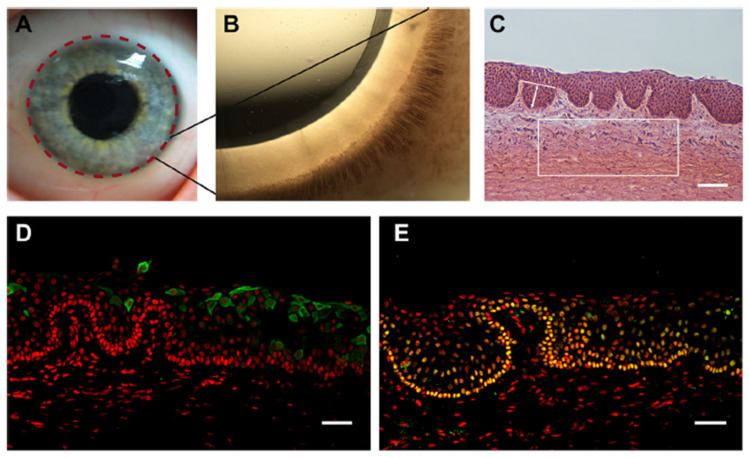
Figure 3.
The human limbal stem cell niche. (A) Describes the location of the limbus with dashed lines on the human ocular surface. (B) Shows a highly pigmented Palisades of Vogt that is visible in the limbus of human. (C) Indicates H and E-stained tangential section of the human limbus, showing the LCs (limbal crypts). The box indicates a representative area of 0.1 mm2 of the limbal stroma. The white line indicates an example of LC width measurements and arrowed line LC depth measurements. (D) CK3- and (E) p63a (green)-stained cryosections of human LCs counterstained with PI (red). Scale bars C: 100 µm, D and E: 50 µm [39].

