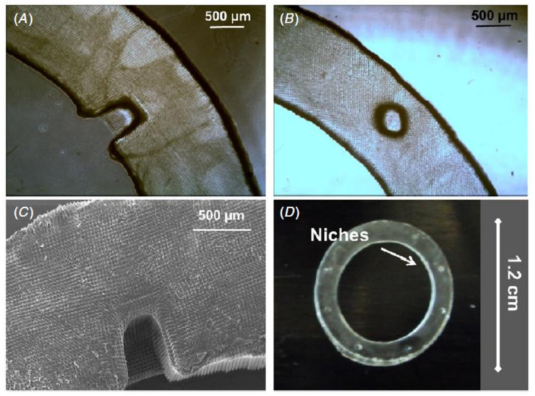
Figure 10.
Phase contrast microscopy and scanning electron microscopy were used to investigate the structure of the PEGDA rings. (A) Describes the optical micrographs of the PEGDA outer ring with horseshoe morphology. (B) Describes the circular morphology of the micrographs of PEGDA. (C) Describes a SEM micrography of the PEGDA outer ring with horseshoe niches. (D) Shows a PEGDA outer ring of diameter 1.2 cm with well-defined artificial micro-pockets of diameter around 300 µm [206].

