
FIGURE 2.
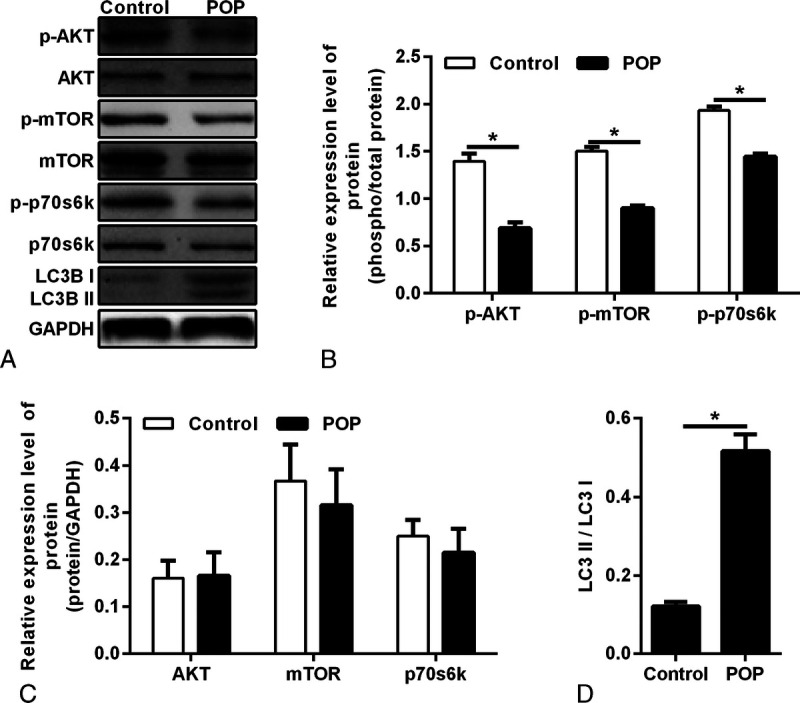
A, Protein expressions of AKT, p-AKT, mTOR, p-mTOR, p70S6K, and p-p70S6K were qualified by Western blotting in vaginal fibroblasts. B, Densitometric analysis for p-AKT, p-mTOR, and p-p70S6K. *P < 0.05. Bar, mean ± SD. C, The relative expression of AKT, mTOR, and p70S6K, which was normalized to GAPDH. D, Densitometric analysis for LC3II/LC3I, *P < 0.05. POP, pelvic organ prolapse.