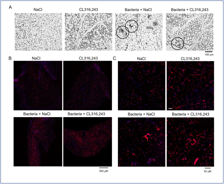
Fig 6. Histological characterization of scWAT infiltration by immune cells.
(A) Representative pictures of four haematoxylin-eosin staining of sub-cutaneous white adipose (scWAT) tissue of mice treated or not with CL316,243 (1 mg/kg/day; 1 week) and infected 48 hours with E. coli. Typical figures of immune cell infiltration and crown structure are black circled. (B, C) Low and high magnification of F4/80 immunostaining (in red) in scWAT of the same mice. Scale bars are indicated.

