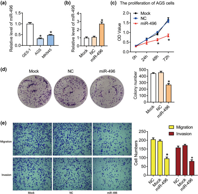
Figure 1.
miR-496 inhibited the proliferation and metastasis in gastric cancer cells. (a) QPCR was performed to detect the level of miR-496 in AGS cells. (b) The level of miR-496 in AGS cells transfected with miR-496 mimics was detected by qPCR. Relative level of miR-496 was analyzed using 2−ΔΔCt method and normalized to mock group. (c) The proliferation of AGS cells was determined using CCK8 assay. OD value (450 nm) was measured every 24 h. (d) Clonogenic assay was used to detect the proliferation of AGS cells. Colony number was counted 2 weeks after the culture. (e) The migration and invasion of AGS cells were detected by transwell assay after the transfection for 24 h. NC = negative control. *P < 0.05.

