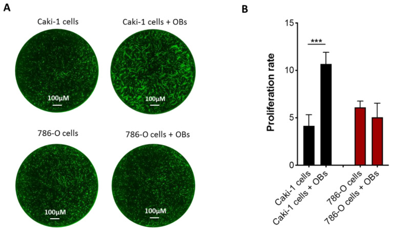
Figure 2.
Effects of OBs on metastatic RCC cell proliferation. (A) Representative images of Caki-1 GFP+ (green) and 786-O GFP+ cells (green) cultured alone or with OBs. Scale bar: 100 μM and magnification 4×. (B) Proliferation rate analysis of Caki-1 GFP+ and 786-O GFP+ cells in a monoculture or cocultured with OBs. Data are expressed as the mean ± SD; *** p < 0.001.

