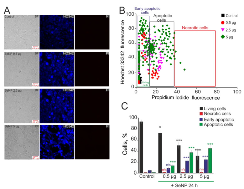
Figure 6.
The effect of different concentrations of SeNP on induction of apoptosis and necrosis in the A-172 cells. (A) Double staining of cells with Hoechst 33,342 (HO342) and Propidium iodide (PI) after incubation with SeNP. (B) Cytogram of the viability of A-172 cells. X-axis—the intensity of PI fluorescence; Y-axis—the intensity of Hoechst 33,342 fluorescence. Cells were stained with the probes after 24 h of SeNP application. (C) The percentage of living cells (black column) and cells in which the processes of early apoptosis (violet column), apoptosis (green column), and necrosis (red column) were detected after incubation with SeNP. The differences between the control and experimental groups are marked with asterisks—* p < 0.05 and *** p < 0.001. n/s—differences insignificant.

