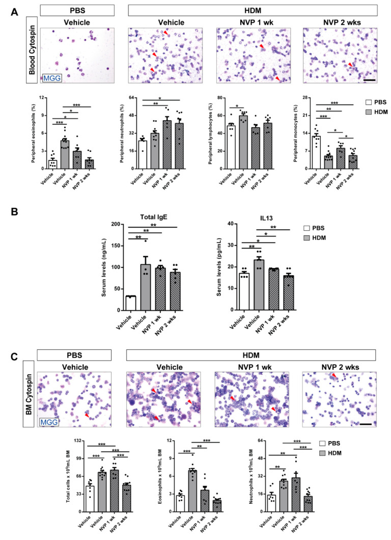
Figure 2.
Pharmacological blockade of IGF1R depletes eosinophil presence in peripheral blood and bone marrow and attenuates the increase in serum IL13 levels after HDM exposure. (A,C) Representative images showing May-Grünwald/Giemsa (MGG) stained peripheral blood and bone marrow cytospin preparations (red arrowheads indicate eosinophils), and differential cell counts for eosinophils, neutrophils, lymphocytes and monocytes in peripheral blood (A), and total cells, eosinophils and neutrophils in bone marrow (C) from HDM-challenged mice treated with NVP vs. controls (n = 7–10 mice per group; scale bars: 50 µm). (B) Total serum IgE and IL13 levels from HDM-challenged mice treated with NVP vs. controls (n = 5–7 mice per group). Data are expressed as mean ± SEM. * p < 0.05; ** p < 0.01; *** p < 0.001 (Mann–Whitney U test or Student´s t-test for comparing two groups and Kruskal–Wallis test or ANOVA multiple comparison test for grouped or multivariate analysis).

