Figure 3.
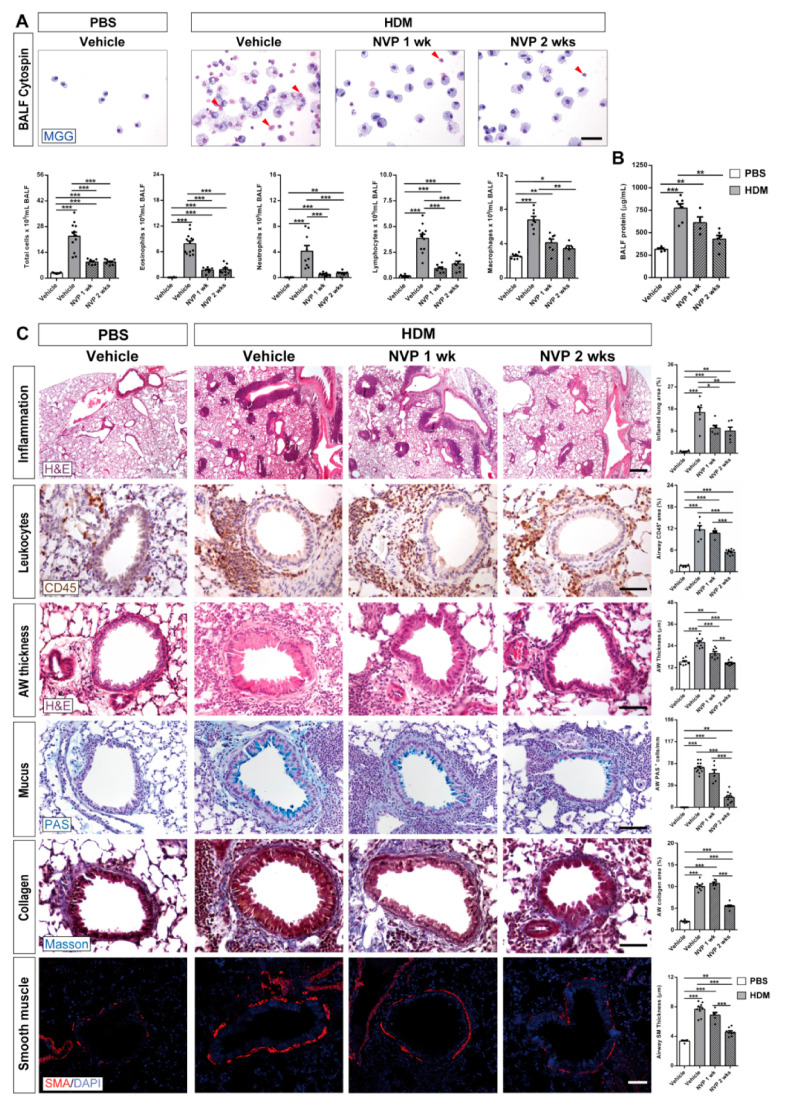
Pharmacological blockade of IGF1R attenuates pulmonary pathology after HDM induced allergy. (A) Representative images showing May-Grünwald/Giemsa (MGG) stained BALF cytospin preparations (red arrowheads indicate eosinophils), and total and differential BALF cell counts for eosinophils, neutrophils, lymphocytes and macrophages in HDM-challenged mice treated with NVP vs. controls (n = 7–12 mice per group; scale bar: 50 µm). (B) Total protein concentration in BALF of HDM-challenged mice treated with NVP vs. controls (n = 5–8 mice per group). (C) Representative images of lung inflammation and histopathology of the proximal airways, and respective quantifications of inflamed lung areas (%) (H&E), presence of peribronchiolar CD45+ area (leukocytes) (%) (brown), airway (AW) epithelium thickness (H&E), number of airway PAS+ cells (mucus-producing cells) (blue), peribronchiolar airway collagen content (%) (Masson in blue) and airway smooth muscle (SM) thickness (SMA in red). These parameters were measured in lung sections from HDM-challenged mice treated with NVP vs. controls (n = 6–10 mice per group; scale bars: 50 µm except for the inflammation panel (400 µm)). Quantifications were performed in five different fields in a random way. Data are expressed as mean ± SEM. * p < 0.05; ** p < 0.01; *** p < 0.001 (Mann–Whitney U test or Student´s t-test for comparing two groups and Kruskal–Wallis test or ANOVA multiple comparison test for grouped or multivariate analysis).

